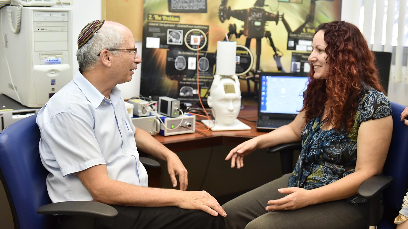
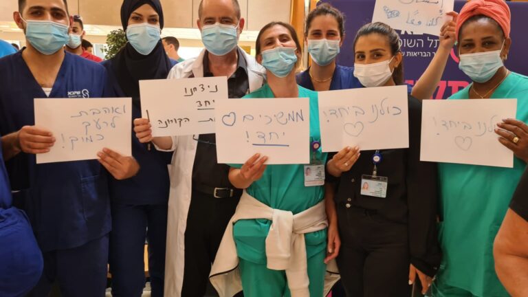
Israeli researchers triumphed over 11 other international teams vying for top nods at the Surgical Robotic Challenge 2016 in London, UK, with their robot for minimally invasive neurosurgery.
The robot, intended for the removal of brain tumors of up to 6-cm in size, is operated through a small keyhole in the skull using laser irradiation and tumor extraction.
The device is composed of a needle assembly: a rigid outer needle and a self-reassembled inner needle. The outer needle is responsible for rotational movement and vertical movement into the tumor, while the inner needle is able to move laterally.
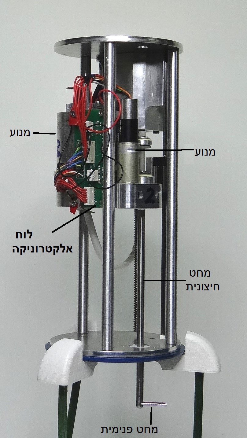
“This project involved many challenges,” says Technion doctoral student Hadas Ziso. “Besides the challenge of miniaturizing the detection and treatment tool, we had to allow the passage of a 90-degree curve in order to minimize the outer needle diameter. For this purpose we developed an inner needle that is flexible enough to pass through the curve, but also strong enough to lead the diagnostic and treatment tool to the tumor accurately, while bearing lateral loads resulted from heterogeneous environment.
“The inner needle mechanism that we developed is based on a chain of tiny magnetic beads, that are partially separated and self-reassembled while passing through a minimal curvature path, Kevlar fibers (a composite material) that pull the mechanism inward, stainless steel links that hold the optical fibers and suction tube, and a polyurethane cover.”
The robot was developed by Ziso, supervised by Professor Moshe Shoham, head of the Medical Robotics Laboratory at the Faculty of Mechanical Engineering, and Professor Menashe Zaaroor, faculty member of the Rappaport Faculty of Medicine and Director of the Department of Neurosurgery at Rambam. The robot is protected by a patent registered in the names of the three researchers and its first inventor, Assistant Professor David Zarrouk, who worked on the project in its early stages during his PhD at the Technion.
The robotic treatment includes several preliminary stages. First, prior to surgery, MRI scans are performed, and the physician prepares the treatment plan on the MR images. Second, a few hours before surgery, the patient drinks a fluorescent medium (5-ALA) that accumulates in the tumor during surgery, so that the robot will rely on both the preliminary MR scans and the morphology of the tumor in real time.
During surgery, ultraviolet (UV) light is projected at the tumor via optical fibers, causing the emission of red light from the fluorescent medium, accumulated within the tumor. The red light allows accurate identification of the cancerous tissue in real time. Based on the information obtained from the detection tool, a high intensety laser is activated, projected from the tip of an optical fiber on the tumor in close range and ablates the tissue.
During treatment, the real time detection is constantly activated to prevent damage to healthy tissue.
Two Israeli companies are involved in the development process of the robot: Prizmatix, which built the optical detection system, and Civan Advanced Technologies, which built the laser system.


