How the brain is wired has long been a question researchers would love to solve. Now, an international ensemble of scientists and engineers has digitalized a piece of the neocortex of the brain of young rats and offers a first-ever inside view of how the brain works.
The Blue Brain Project, a key part of the European Union’s 10-year Human Brain Project, has released a detailed computer representation of the microcircuitry of a small area of the rat’s somatosensory cortex, the part of the brain responsible for the sense of touch.
Using supercomputers, the 82 Blue Brain Project engineers and scientists — from institutions in Israel, Spain, Hungary, US, China, Switzerland, Sweden and the UK — simulated electrical behavior of virtual brain tissue and found that while some of the behavior observed in previous brain experiments was a match, other simulations revealed novel insights into the functioning of the neocortex.
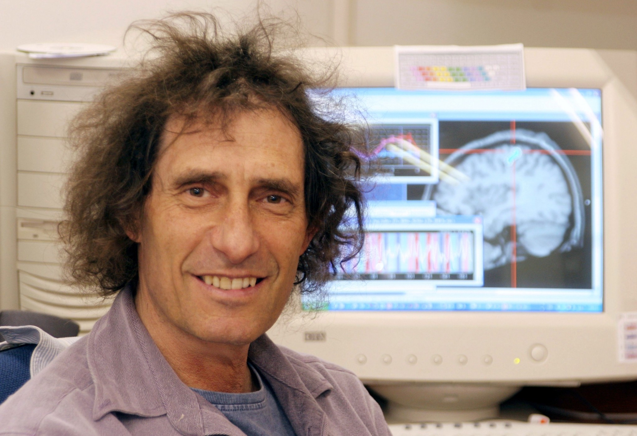
“With the Blue Brain Project, we are creating a digital reconstruction of the brain and using supercomputer simulations of its electrical behavior to reveal a variety of brain states. This allows us to examine brain phenomena within a purely digital environment and conduct experiments previously only possible using biological tissue,” said Hebrew University of Jerusalem’s Prof. Idan Segev, a senior author of the research paper, recently published in the journal Cell.

The findings culminate 20 years of biological experimentation that generated the core dataset, and 10 years of computational science work to develop the algorithms and build the software ecosystem required to digitally reconstruct and simulate the tissue.
“The insights we gather from these experiments will help us to understand normal and abnormal brain states, and in the future may have the potential to help us develop new avenues for treating brain disorders,” said Segev.
Supercomputers to the rescue
While science is still a long way from digitizing the whole brain, the study proves that it is feasible to digitally reconstruct and simulate brain tissue and to reveal novel insights into brain function.
Segev, a member of the Hebrew University’s Edmond and Lily Safra Center for Brain Sciences and director of the university’s department of neurobiology, sees the research as building on the pioneering work of the Spanish anatomist Ramon y Cajal more than 100 years ago.
“Ramon y Cajal began drawing every type of neuron in the brain by hand. He even drew in arrows to describe how he thought the information was flowing from one neuron to the next. Today, we are doing what Cajal would be doing with the tools of the day: building a digital representation of the neurons and synapses, and simulating the flow of information between neurons on supercomputers,” said Segev.
“The insights we gather from these experiments will help us to understand normal and abnormal brain states, and in the future may have the potential to help us develop new avenues for treating brain disorders.”
“The digitization of the tissue is open to the community and allows the data and the models to be preserved and reused for future generations.”
Supercomputers are an important new tool for neuroscience.
“Building the digital reconstructions, running the simulations and analyzing the results required a supercomputing infrastructure and a large ecosystem of software. It was only with this kind of infrastructure that we could solve the billions of equations needed to simulate each 25-microsecond time-step in the simulation,” said Felix Schürmann, a senior author affiliated with École polytechnique fédérale de Lausanne in Geneva, and head of the project’s software development team.
A major milestone
The computer reconstruction depicts the shapes and electrical behaviors of approximately 30,000 nerve cells (neurons) in the reconstructed tissue, and approximately 40 million synapses (the sites of connection and communication between neurons) that they form.
“The reconstruction is a first draft; it is not complete and it is not yet a perfect digital replica of the biological tissue,” said Henry Markram, a professor at the École Polytechnique Fédérale de Lausanne and director of both the Blue Brain Project and the Human Brain Project.
The researchers performed tens of thousands of experiments on neurons and synapses in the neocortex of young rats and catalogued each type of neuron and synapse they found. The resulting data collection is one of the most comprehensive to date on a part of the brain.
To complete the microcircuitry map, the researchers developed software to mine published findings from laboratories worldwide, and algorithms that use the available data as landmarks.
“We can’t and don’t have to measure everything,” said Markram, who received his graduate degree from the Weizmann Institute of Science in Rehovot. “The brain is a well-ordered structure, so once you begin to understand the order at the microscopic level, you can start to predict much of the missing data.”
Now other scientists will be able to use the Blue Brain data and reconstruction to test theories of brain function.
“The job of reconstructing and simulating the brain is a large-scale collaborative one, and the work has only just begun. The Human Brain Project represents the kind of collaboration that is required,” said Sean Hill, a senior author.


